I met Hickory when we leased her as a broodmare from a nearby rancher. She arrived on our farm in southwestern Virginia on a warm spring day. Right away I was impressed by how easily she handled, how calm she was and the silkiness of her buckskin coat.
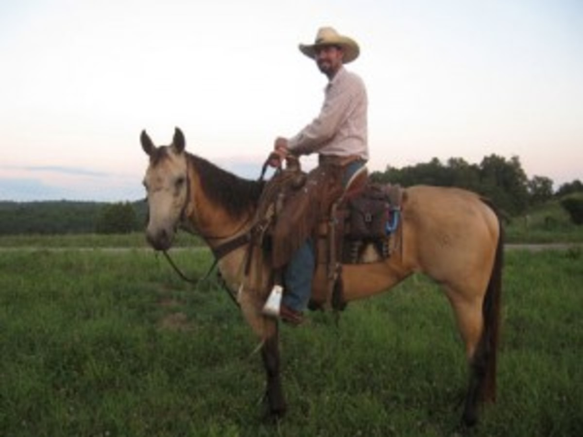
But I also noticed a large scar that snaked across and down her face. She had obviously been seriously injured at some point. Perhaps, I thought, she reared in a trailer and smashed her face on the roof. But she didn’t have the slightest aversion to trailer loading, or anything else for that matter. We asked her owner about it, but he had no answers.
Hickory bred easily and gave birth to a fantastic little filly on Easter morning, 2009. The original plan called for Hickory to go home once the baby was weaned, but her owner hoped to find her a new home instead. We didn’t need another mare, but such a wonderful horse deserves a great home, so we arranged a deal: Our neighbor, who uses horses to work cattle, would take her.
Hickory quickly picked up her training under saddle and proved to be talented at sorting and roping. She was willing, trusting and fun to ride, and she spent the occasional weekend competing in local rodeos. Also intrigued by the mare’s scar, our neighbor researched her history and found her breeders. With a phone call, he heard the amazing story of the injury behind the scar. Here it is.
A horrific sight
Jon and Marywade Gilbert were at the local fairground in April of 2006 when their head ranch hand called in a panic. “I couldn’t make out much of what he was saying, but it was obvious something terrible had happened,” says Jon Gilbert. “I was running an auction, so Marywade left immediatelyfor home.”
Home was an operation called HorseBreakers Unlimited in Dewey, Arizona, where the Gilberts breed Quarter Horses. When she arrived, Marywade found that MJG Scottish Hickory, a 3-year-old buckskin filly, had crushed the front of her face.
“We had what we call out here a microburst,'” Marywade Gilbert explains. “It’s a very strong downward blast of wind, and it snapped a tree off next to her paddock. It fell and hit the fence and scared both of the fillies that were there. They took off and ran. The other filly fell and slid under the fence, and Hickory ran to the corner.”
Unfortunately, the panicked filly didn’t stop or jump when she reached the pipe fence. “She hit the corner post, which is set in concrete, just below her eyes,” Marywade Gilbert says. “On the right side of her face [from her forehead to her muzzle], there was just kind of nothing there except her eye and her nostril—everything else was just broken bone and open skin. The hole on the right side of her face … that’s what she was actually breathing through. My first thought was, ‘There’s no way a horse can survive this.'”
Despite the horrible injury, the filly was remarkably quiet and appeared to be getting adequate air. Marywade called her veterinarian, James Bleak, DVM, of Central Arizona Equine in Camp Verde, who arrived quickly. After a quick assessment of the wound, he delivered some news that shocked the filly’s owner:
“I told her that I thought Hickory would be fine,” says Bleak. “The injury was horrific looking, but no vital structures were damaged.” Head injuries in people are serious because of the risk of brain involvement, but the equine brain is much smaller and sits above the eyes, between the ears and in front of the poll. Hickory had smashed the front of her face, crushing the thin layer of bone that lies over her sinus cavities, but she had not injured her eyes, brain, teeth or other critical structures.
“Properly treated face and head injuries in horses tend to heal fairly well,” Bleak says, “even when they start out looking gruesome.” He continued his exam to assess the best approach for repairing the damage.
“She had split her forehead open. We had a skin flap there that was probably four inches long on the vertical side, and maybe two to three inches on the top horizontal side,” Bleak says. “Right on the forehead, we had multiple fractures that extended down the bridge of the nose a little bit further. It had peeled that skin back and we could definitely see what was going on. And I could even feel under a portion of the skin that wasn’t lacerated that the bone had slid under another part of bone there.” The interior of the filly’s airways was exposed, and one of the bones forming the orbit of her right eye was also fractured.
The injury was complex enough that Bleak didn’t think it wise to begin a lengthy procedure in waning light at the farm with the filly only under sedation. He suggested sending Hickory to Chaparral Veterinary Medical Center in Cave Creek, about two hours away. There, not only could the filly be treated in a fully equipped surgical facility under general anesthesia, but a veterinarian on staff, Renee Andrea, DVM, Dipl ACVS, specializes in equine facial reconstruction.
The Gilberts agreed, and Bleak called the clinic to confer with Andrea. The two veterinarians decided it was best to stabilize Hickory where she was with painkillers, start her on antibiotics and then trailer her to the clinic the following morning for surgery.
After administering the medications to Hickory, Bleak applied temporary sutures to the large wound on her forehead. “Damaged skin will shrink over time,” he explains. “If we just left the wound open until the morning, she wouldn’t have had sufficient skin left to recover the area. I pulled the edges shut and sewed them with large sutures. It wasn’t pretty, but they were only intended to hold overnight and come out the next day.”
Assembling the pieces
Andrea was waiting when Hickory and Marywade Gilbert arrived at Chaparral the next morning. “When I first saw her I could tell it was really, really bad,” she says. “But I also knew I could fix it. If you have an owner with the financial means to pursue reconstructive surgery and the patience to do any necessary follow-up procedures, it’s amazing what you can restore.”
After removing the temporary sutures, Andrea peeled back the open skin to see the extent of the damage: “The bones of the front of her face were broken in multiple places and pushed back into her sinus cavity. The bone
under her eye, part of the orbit, was also fractured and out of position, but the eye itself was in good shape.”
First, the surgeon used forceps to return the orbit to its normal position. Then she searched through the exposed sinuses to remove every piece of loose bone she could find: “It’s kind of like putting a jigsaw puzzle back together, but you have to find all the pieces first.”
However, not every bone fragment would be replaced. “In a situation like this, I have to look at each fragment carefully,” Andrea says. “If it’s discolored around the edges or otherwise looks like it will die, I don’t replace it.” The search was important for another reason as well: The sinuses are naturally full of bacteria, and a fragment of dead tissue left in the cavity could trigger a serious infection.
With the bone fragments retrieved and sorted, Andrea began reassembling Hickory’s skull. First she repositioned large sections of the frontal (forehead) and maxillary (overlying the upper cheek teeth) bones that had been pushed under each other and inserted small wires to hold them in place. Then she wired in smaller fragments in their proper positions.
Areas in the skull where bone fragments could not be replaced were left open. “Normally, bone wouldn’t grow back in these places, and the skin might not cover it, so the horse has a blowhole of sorts on his face,” says Andrea. One option is to cover the openings with metal plates. But, she says, “I don’t use plates on skull repairs because the bone is so thin.” She does, however, use another technique to cover the holes: “It’s really like my secret weapon in these cases.”
Andrea made a large incision through the skin in an area adjacent to the wound site, exposing healthy, intact skull bone. Then she peeled back the thin membrane that covers the bone, called the periosteum, creating a loose flap still attached to its original blood supply. Carefully, she repositioned the flap over the reconstructed bone.
Among other functions, the periosteum contains specialized cells called osteoblasts that generate new bone cells to aid in repairing fractures. “The periosteum has wonderful healing factors in it,” Andrea says. “So much so that areas below it will grow new bone to fill any gaps in the reconstructed skull.” The healthy bone the periosteum was removed from isn’t adversely affected by its loss, she adds.
With the bones repositioned and secured and the periosteum flap in place, Andrea began the painstaking process of suturing the skin closed over Hickory’s face. “I used very tiny subcutaneous sutures that would dissolve over time and not have to be removed,” says Andrea. “That is really intricate work, but I find it gives the best cosmetic results.”
After nearly six hours of surgery, the reconstruction of Hickory’s skull was complete. But the team wasn’t done yet. “As horses come out of anesthesia they can panic, lose their balance and flail around,” says Andrea. “After surgery like this, one slight knock of the head against a wall and all of your work is destroyed.”
To protect Hickory’s head, the surgical team constructed a large “helmet” from thick Styrofoam, securing it to areas of her skull that had not been damaged. They also used a head and tail rope to help steady the filly as she rose to her feet. But true to her calm nature, Hickory recovered without incident and returned home three days later, still wearing her protective headgear.
Bleak made several visits to check on Hickory as she recovered. “Mainly what you worry about in these cases is infection,” he says. “But we had started antibiotics within hours of the injury, and the surgery cleaned out anything that could have brewed problems. I watched carefully for nasal discharge, a smell or a fever that might indicate infection, but that never happened.”
Hickory looked a bit like Frankenstein’s monster in the weeks after her surgery, but eventually her skin healed, the sutures dissolved and the scars began to fade. The contours of her face were nearly normal and her breathing was strong and silent, all signs that the bones had knitted back together as expected.
Six months later, Hickory was a normal, healthy mare who just happened to have a few scars on her face, and the Gilberts decided there was no reason she couldn’t become a useful working horse. Their plan had always been to sell her, and so that’s what they did—starting her on a path that led to her new life on the ranch next door to mine.