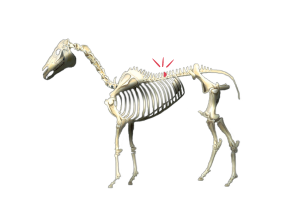
Overriding Spinous Process, otherwise known as “kissing spine,” can cause back pain and poor performance, especially when two or more vertebrae touch or overlap.
A November 27 press release from Canada’s Equine Guelph discusses a new approach for treating this condition. Earlier this year, Assistant Professor Dr. Nathalie Cote from the department of Large Animal Surgery at Ontario Veterinary College presented a new, less invasive surgical option to treat this issue that is showing great preliminary results.
Where and why
While there is very little movement in the lumbar area of a horse’s spine, the thoracic area just in front of it has a slightly wider range. Thoracic vertebrae allow side-to-side flexion, a little rotation, and flexion and extension, which allow the back to move up and down. Impingement most frequently occurs under the saddle area between thoracic vertebrae T13 to T18 with T14 to 16 (right where the rider sits) being the most common.
Not all riding horses with kissing spine will present with clinical signs. In fact, it is not uncommon to find kissing spine post-mortem in riding horses that have not shown obvious signs of pain. Kissing spine has also been found in post-mortems of extinct and undomesticated horses, which leads to conclusions that in some cases conformation plays a role.
Four grades of impingement
In horses with spinal impingement that do present with back pain and behavioral signs (bucking, rearing, refusing jumps, being girthy or sensitive to brush, etc.), there are typically more than one vertebra affected, and the severity of impingement is greater.
A grading system of one to four is assigned when diagnostic radiographs are performed. In grade one, there is narrowing of the space between the spinous process, and in grade two, that loss of space is significant. In grade three, bone remodeling has begun, and in grade four, malformation will be significant, and the space is almost impossible to go in between in past surgical options.
A quicker, less invasive treatment
As Cote explained, desmotomy (where ligaments are severed and potentially redistributed) with a scissor tool proves challenging when it comes to grade four cases, and treatments involving partial removal are costly and invasive.

The modified procedure of surgical desmotomy of the interspinous ligament uses a tool much smaller than scissors, allowing a smaller incision to be made and resulting in improved cosmetic appearance. The modified procedure makes it easier to work with the minimal spaces seen in grade four cases.
It is a quicker, less invasive procedure that begins with inserting needles into the interspinous spaces. X-rays are then taken, and the needles are removed for the areas that will not be operated on and left in for those that will be worked on. A one-centimeter incision is made using a narrow tenotome tool to dissect the identified ligaments which restores space between the thoracic vertebrae.
Good prognosis
So far, clients adhering to a six-week rehabilitation program of stall rest and hand-walking (three weeks) followed by lunging, core strengthening and mobilizing exercises have generally received a very good prognosis for their horses’ scar tissue to heal well and mobility to be restored. Some clients reported their horses going back into full training as early as four months after the surgery.
Cote presented her findings on the modified procedure of surgical desmotomy of the interspinous ligament to a group of Ontario Association of Equine Practitioners (OAEP) on February 15, 2023, at the University of Guelph. Here is her presentation.
Equine Guelph supports a number of high-quality projects at the University of Guelph, by virtue of funding provided largely by the racing industry (Standardbred, Thoroughbred and Quarter Horse organizations): the Horse Improvement Program from the Horsemen’s Benevolent and Protective Association and the E.P. Taylor Foundation, started by veterinarians in the Thoroughbred industry, and now maintained in trust by the University and Equine Guelph.

