As a top-level hunter-jumper, Charlie had always received top-notch care, and he had been sound and injury free through several years of competition. So it was a surprise to everyone when the 12-year-old warmblood gelding walked out of his stall dramatically lame in his right front limb one June morning.
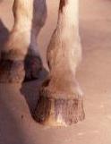
“He was moderately lame with heat and diffuse from his fetlock to his coronet band,” says Rachel Buchholz, DVM, of Northwest Equine Performance in Mulino, Oregon.
A hoof abscess is usually the first diagnostic consideration when a severe lameness develops so suddenly. “Frankly, you kind of cross your fingers that it’s an abscess,” says Buchholz. “Abscesses are one of the few things that can cause sudden dramatic lameness in a hoof, with that type of swelling, that isn’t a broken bone or something just as devastating.”
Hoof abscesses are pockets of pus that form within the hoof capsule after bacteria enter through a defect, such as a crack in the sole. Although abscesses are acutely painful, they do no long-term damage. Once the pus is drained—either the fluids will work their way to the sole, heel bulb or coronary band and rupture on their own, or a farrier or veterinarian may pare into the sole to provide an outlet—the damaged tissue heals and the horse returns to full soundness.
Buchholz performed a full lameness diagnostic workup to look for the source of Charlie’s hoof pain. This external examination involves watching how the horse bears weight as he moves, palpating the structures of the leg and foot, hyperflexing joints to accentuate any lameness, looking for abnormalities in the hoof wall or sole, using hoof testers to locate specific areas of sensitivity within the foot, and administering nerve blocks to deaden pain to pinpoint a source of lameness. “He was responsive to heel bulb palpation and had mildly elevated digital pulses in that foot,” she says. “When we blocked his foot with a simple heel block, he was much sounder.”
Still suspecting an abscess, the veterinarian pulled the gelding’s shoe to look for any signs of bruising or trauma to the sole. “He had pads, so we pulled those off also,” she says, “but we didn’t find anything remarkable underneath them. Then we took x-rays, but they were just as unexciting.” The radiographs showed no broken bones, which was of course good news, but Buchholz still had very few clues.
Charlie’s owners were faced with two choices. “We have lots of great diagnostic tools in the clinic, so we could have gone looking further right then,” says Buchholz. “But that can be expensive and isn’t always necessary. The other option was taking him home and soaking his foot for a few days to see if the abscess would burst.”
The decision was to take Charlie home to see what might develop.
Looking deeper
Two weeks later, Charlie’s lameness had diminished only slightly, and there were no signs of an abscess breaking through the hoof. “He had been on stall rest and was having his hoof soaked twice a day,” says Buchholz. “If an abscess hasn’t shown itself by then, chances are that’s not what you’re dealing with.”
Charlie returned to the clinic for further diagnostic work. Another round of flexion tests, hoof testers and blocks turned up no new findings. The next step would be some sort of high-tech imaging to try to get a better look at the interior structures of the hoof. “We talked about ultrasound but, frankly, with the swelling he still had, and the likelihood of the issue being deep in the foot, we doubted our ability to see much,” says Buchholz.
A more useful diagnostic imaging option, she advised Charlie’s owners, might be magnetic resonance imaging (MRI). For an MRI, the body part to be scanned is placed in a strong magnetic field, which aligns the hydrogen atoms of every water molecule in the tissues in one direction. Then a second electromagnetic field, perpendicular to the first, pulses on and off, pulling the molecules out of alignment and releasing them to fall back in line with the first magnetic field. As the molecules oscillate between the two magnetic fields, they release detectable radiofrequency signals. And because the water molecules in tissues of different densities respond to the pulses at different rates, a detector can use these signals to create images of every internal structure, including both hard and soft tissues.
The very clearest MRI images come from high-field machines. But these require that the horse be put under general anesthesia and placed on a specialized gurney so that the leg can be rolled into a fully enclosed, horizontal chamber. Low-field MRI machines have weaker magnetic fields, but when used on the lower limb—and the hoof especially—they can produce images nearly equal in clarity to a high-field machine. The advantage of low-field machines is that they can be used vertically, which means that a horse’s leg and hoof can be imaged while he is standing under sedation.
“The level of sedation is less than we’d use for a dental visit, which is safe and easy to manage,” says Buchholz. “We could do it right away.” The only alternative was to continue Charlie’s stall rest, with anti-inflammatory medications, to see if the still-undiagnosed injury would heal on its own. Charlie’s owners opted for the standing MRI.
With Charlie sedated and positioned properly, the veterinary team started the procedure. “I am usually the one running the MRI,” says Buchholz, “so I’m right there looking at the images as they come up. Sometimes I can see right away what’s going on, but we will also send them out to a board-certified radiologist for a final opinion if we aren’t sure.”
A clear answer
Charlie’s problem was instantly apparent: The lateral coffin collateral ligament in his right hoof was bright white—the result of extensive fluid accumulation. “What we saw was indicative of what we call ‘extensive fiber disruption,’” says Buchholz. “His ligament wasn’t torn through, but it was pretty darn close.”
The collateral ligaments—two per hoof—connect the short pastern bone to the coffin bone. “They are big, supportive structures that help keep the bones in a stable position and aligned with each other,” says Buchholz. These ligaments can be damaged from the twisting forces of a sudden turn or a slip on uneven footing. “It doesn’t usually happen from overuse or extreme activities,” she adds. “It’s typically just rotten luck.”
With a definitive diagnosis, Buchholz was able to develop a treatment plan targeted to Charlie’s specific problem: “For injuries like this, we typically recommend a two-pronged approach.” First, the veterinarians would use extracorporeal shock wave therapy (ESWT) to direct high-intensity shock waves into the injury site. “This reminds the body that there’s an injury there and creates a bit of an inflammatory response,” she says.
Next, they would inject platelet-rich plasma (PRP) directly into the injured ligament. PRP involves drawing a sample of a horse’s own blood and running it through a specialized centrifuge to separate out the platelets, the small blood cells that play an essential role in healing injuries. When they encounter injured tissues, platelets are well known for forming clots or scabs, but they also release a number of growth factors, substances that stimulate the regrowth of healthy tissue. When the platelets and a small amount of plasma are separated from the rest of the blood sample for PRP therapy, the mixture is injected directly into the injured soft tissue.
“We usually do the shock wave and PRP on the same day—one right after the other, in that order,” says Buchholz.
“I explain to people that we are creating an inflammatory response with the shock wave. Then, as the body is mounting a healing response, we inject the PRP to give it more ammunition.”
After this treatment, Charlie was sent home for more stall rest, shoeing changes and controlled hand-walking.
Rest and recovery
The gelding was returned to the clinic for a recheck almost two months later, in August. “He was sound at that point, nonresponsive to palpation or hoof testers, and the swelling on his leg was much smaller and firmer,” says Buchholz.
With the swelling reduced, the veterinary team used ultrasound to examine the healing ligament, and the images suggested that everything was going well. “Ligaments never look 100 percent like they did pre-injury,” Buchholz says. “But when they are healing well, you will see much less swelling.”
Charlie was cleared to start walking under saddle for 20 minutes a day, increasing to 60 minutes over several weeks. “Cautious rehab after an injury like this is very important, especially for older horses,” Buchholz says. “You want to start adding stresses to the area to see how it responds, but you don’t want to push it too far.”
A horse who goes lame during the rehabilitation is less likely to ever make a full recovery, says Buchholz: “Sometimes a horse will get an entirely new injury in the same limb. It’s important to do a diagnostic workup again to see exactly what you’re dealing with every time, even when you think you know. Not all horses read the book.”
If, however, the exam reveals that reinjury has occurred, expectations for recovery need to be adjusted, she adds: “Typically if the horse reinjures the same area multiple times he’s going to need a different job. The prognosis for returning to what he used to do goes down significantly with each reinjury.”
Charlie remained sound throughout his convalescence. “We did another clinical recheck in November, followed by a clinical and MRI recheck in January,” says Buchholz. The image showed a dramatic improvement: “You could tell where he had been injured, but this looked as good as we could hope for.”
After that, Charlie was allowed to begin trot work, only five minutes at a time to start, adding five minutes per week as long as he remained sound. When he was up to 20 minutes of trotting with no difficulties, he was rechecked again and cleared to start cantering and slowly work up to jumping small fences. “We didn’t know if he’d ever go back to flat work, much less jumping,” says Buchholz, “but at our last clinical check, a year and a month after the original injury, he was doing great.”
Charlie’s story illustrates how integral an accurate diagnosis is to a horse’s recovery, Buchholz says: “Without knowing exactly what we were dealing with, all we could do was rest the horse and hope it worked. MRI and other high-tech diagnostics may seem expensive on the outset, but a lot of times people will spend six or eight months trying to figure out what’s going on, and they spend the same amount in the end and may or may not have an answer. Once you know what’s going on, you can focus your efforts and resources on treatment and, hopefully, start seeing results.”
This article first appeared in EQUUS issue #440.