At first Ashley Jenkins thought her 19-year-old mare, Belle, had a respiratory infection. “She had a slight trickle of yellow fluid coming from her left nostril,” Jenkins says. “I assumed she had a cold.” A cold wouldn’t have been surprising, given the weather. The winter had been uncharacteristically long and harsh for Virginia, and by March 2014, a warm spell followed by another plummet in the temperatures had left everyone feeling a little out of sorts.
But then, says Jenkins, “I noticed she also had a large lump under her left eye.”
Jenkins had owned Belle for 16 years and had shown the mare under the name Skip Eagles Star in American Quarter Horse Association-recognized competitions and all-around and hunter-under-saddle classes. Now Belle was retired to Jenkins’ parents’ farm. Apart from a bout of tying up when she was younger, the bay mare had always been healthy. “I still ride her occasionally,” says Jenkins. “But she’s mostly a pretty pasture ornament.”
Despite the runny nose and the odd lump on her face, Belle seemed happy and comfortable. She was eating well and showed no signs of pain, distress or illness. Nevertheless, Jenkins called Chris Robertson, DVM, of Blue Mountain Equine, to investigate.
Robertson immediately had a bad feeling about the lump on Belle’s face. The swelling had a low profile, but it covered a wide area. “It had the contours of a baseball, as if it was shoved up into her nose and I could see the top sixth of it,” he says. Even more disturbing was the fact the lump was apparently formed by bone being pushed out from the mare’s face.
“I could tell that the lump we were feeling was actually her skull bulging from the inside out,” says Robertson. “The only thing I could think of that would cause a lump like that on the face was a very large and aggressive tumor within the sinus cavity.” Sinus tumors can be diffi- cult to treat in horses. “Even if you can remove some, you can’t expect them to go away completely,” says Robertson. “They tend to come back and grow until the horse is really uncomfortable. We had seen a horse a few years ago with a fibrosarcoma in his sinus, and it didn’t end well.”
Robertson inserted a small hypodermic needle into the lump on Belle’s face in an attempt to aspirate some fluid. “If a lump is caused by an infection or abscess, you’ll be able to pull stuff out with a needle,” he says, “but you won’t get much if it’s a tumor.” When multiple tries yielded only a very small amount of bloody fluid, the grim diagnosis seemed all the more certain.
As Robertson explained his suspicions to Jenkins, her heart sank. “He said that he was pretty sure it was a tumor and the outlook wasn’t good,” she says. “He also said he could refer us to a large clinic for x-rays and a biopsy to be certain, and that they might attempt to treat it. The situation didn’t sound encouraging at all, but she’s my pride and joy. I didn’t hesitate to ask for the referral.”
Robertson arranged for Belle to be seen by Paul Stephens, DVM, at Blue Ridge Equine Clinic in Earlysville, Virginia, the next day. In the meantime, he started the mare on a course of antibiotics, in case the lump was indeed an infection, along with an anti-inflammatory medication.
But that night, another winter storm rolled through and covered Northern Virginia with nearly a foot of snow and ice. The trip to the clinic would have to be postponed. “We couldn’t safely ship her anywhere,” says Jenkins. “It was very upsetting to be able to do nothing but wait.” Meanwhile, the lump on Belle’s face grew larger.
A game changer
A week later, the roads were finally clear enough to take Belle on the two-hour trip. After examining the mare, Stephens met with Jenkins.
“The lump was just as Robertson had described it to me—very large, focal and very firm,” says Stephens. “In that area of the face, there is very little soft tissue—it’s just the skin stretched over bone—so if you get an infection or an abscess, it tends to be diffuse and spread out, not collected. The only thing I could think of that could grow and mushroom like this, deforming the bone, was a tumor. I was virtually certain that’s what we were dealing with, and I told Ashley that.”
The news was devastating. “Dr. Stephens told me he still had to biopsy the lump to be certain, but he set the stage to give me some very bad news. I called my dad and started crying, then my friends, just sobbing about Belle.”
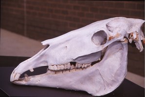
Meanwhile, the veterinary team took a series of radiographs in preparation for a biopsy. The images revealed some unexpected—and game-changing— information: “We saw a dramatic soft- tissue density mass, pretty well centered in the sinus,” Stephens says. “But what was also very obvious was a fractured tooth just below the mass. Not just split, but with several millimeters separation. The entire bony socket that held the tooth was deformed and expanded.”
This was good news, he adds, because it meant that the mass might not be a tumor: “A tumor cannot split a tooth. It might invade the adjacent bone and soft tissue and push the tooth out of position but not cause it to fracture.”
A more likely scenario was that Belle had fractured her tooth first, and that had led to a massive infection after feed became trapped in the crevice. Belle hadn’t shown any other signs of an infection—such as copious nose drainage, bad breath or an elevated temperature—but it remained the best working theory at the moment.
“Our next step was an oral exam,” says Stephens, which confirmed the fracture and the presence of feed in the area. “Then we decided to open the mass from the outside, by cutting into the face to see what was in there.” With Belle heavily sedated, the surgeon made a small incision through the degraded bone directly over the lump and reached in with a forceps to see what he could pull out.
Jenkins had just hung up the phone and was trying to regain her composure when Stephens burst into the waiting area. “He was so excited,” she recalls. “He was saying, ‘It’s feed! It’s not a tumor. There’s feed coming out!’ I was definitely confused, but he seemed really happy.’”
An unusual accumulation
Normally, when a horse’s tooth fractures, the bony socket remains intact. “Bacteria may cross through the bone at the tooth root to form an abscess, and in the last three upper cheek teeth this can result in a sinus infection,” says Stephens. But Belle’s case was different: “When the tooth fractured—however it fractured—it fractured the bone above it as well, in the deepest part of the tooth socket. Feed was pushed right up through it to collect in the sinus cavity above the area. When we cut into the lump, we found it was all packed, rotting feed.”
It’s remarkable that Belle continued to eat normally with such extensive damage to her mouth, but not unheard of, says Stephens: “Horses will adapt to survive. I’ve seen horses with tooth fractures and nasty infections and they’ve never turned down a meal. They just switch to chewing on the other side of their mouths. It’s amazing what they can tolerate.”
Leaving Jenkins to call her family and friends back with the good news, Stephens returned to begin treating Belle. “First we needed to get the pocket cleaned out, working from the outside of her face,” he says. “We spent the next 15 to 20 minutes pulling out the feed with forceps and then flushing the area with saline. We also scraped the inside of the pocket to remove any tissue that was necrotic0. The smell was absolutely horrendous, but eventually we were successful.”
Next, Stephens turned to the inside of Belle’s mouth. “Using a speculum to keep her mouth open, we were able to use dental forceps to remove each half of the tooth,” he says. “I was a bit worried we wouldn’t be able to get the entire tooth out, but eventually we were able to.”
Stephens also removed any bone that looked dead, infected or dying. “When we were done, we had this huge hole going up through the mouth where the tooth had been, directly into the mare’s sinus and then out her face straight to the great outdoors,” he says. “It was dramatic, to say the least.”
Filling the void
Normally, when an upper cheek tooth is removed, the veterinarian fills the socket with an acrylic plug. “This prevents feed material from being packed up into the socket,” says Stephens. “If the socket is shallow, we just pack it for a few days with gauze, like when you get your wisdom teeth pulled, until tissue begins to fill in.” For deeper sockets, the veterinarian may fashion an acrylic plug that fits snugly into the area to accomplish the same goal over a longer period of time.
But in this case, Stephens was going to have to improvise. “Belle certainly had a deep socket, but because it was very large and didn’t taper, it was going to be problematic to get a plug to hold in there,” he says. “But I couldn’t leave it open. What I elected to do was pack the hole from the outside with rolled gauze I’d made into a ball. Then, I packed the socket from the inside of the mouth with sterile gauze. By coming at the space from both sides, we were able to fill it completely.”
This gauze packing would be changed twice daily as the socket began to heal. “As the tissues get healthy, they start to fill in,” says Stephens. “You pack the gauze in tight enough to hold but loose enough to leave room for the new tissues. Eventually, you’re packing less and less into the space.”
Not only would the gauze keep feed out of the socket as Belle ate, but the rough texture of the material would debride the wound each time it was removed. “Necrotic issue will stick to gauze,” says Stephens. “That means that as we changed the packing, we’d be removing any dying tissue that could lead to an infection.” Although Belle was still on an antibiotic, and would be for a few weeks, any extra protection against infection would be beneficial.
Removing and repacking the gauze wasn’t simple, however. “We had to do it with Belle under light sedation,” says Stephens. “We’d remove all the gauze and lavage the socket, from the outside down into her mouth. Then we’d repack the entire area, hopefully with less gauze than we needed before.”
Belle remained at the clinic for another five days, then she was moved to a lay-up facility where Stephens continued to visit daily to tend to her wound. “There wasn’t any way we could do this ourselves at home,” says Jenkins. “To help us with the cost, Dr. Stephens arranged to have her moved to the most amazing facility I’ve ever seen, and then he went out there on his own time and his days off to take care of her.”
Belle’s wound healed quickly. Less than two weeks after the tooth was removed, the socket had closed enough to be fitted with an acrylic plug. Plugging the socket would eliminate the need to pack gauze in the wound from the inside of the mouth and provide more protection against feed contamination and infection. For this procedure, Belle was shipped back to the clinic.
“The plugs are made of two compounds you mix,” Stephens says. “They are moldable for a few minutes, but then they set up and harden. The idea is to shape it to fit snugly just a half-inch deep into the socket from the gum line. You try to mold a flange to fit over the gum on the sides of the empty socket and then a little bit at the base of the tooth in front and the tooth behind, creating a bridge.”
The socket would continue to heal with the plug protecting the gum. “Sometimes the plug falls out in a few weeks, sometimes it’s a few years, and sometimes it never falls out—and that’s OK,” says Stephens.
Aftercare and unanswered questions
With the plug in place, Belle was sent home and returned to Robertson’s care. “I felt comfortable sending her home, but we weren’t out of the woods yet,” says Stephens. “It seemed unlikely that the sinus wouldn’t need additional debridement to remove pockets of residual disease. She was still on an antibiotic, but that wasn’t any guarantee. It was almost too much to hope that it was just going to continue to heal.”
Robertson visited Belle every two days. “Her sinus was still open to the exterior, so I’d remove the gauze, flush the area and repack it,” he says. “After the first week, though, enough tissue had filled in that I didn’t really have room for the gauze. And a few days after that, the exterior wound had closed completely.”
Her mouth had healed, and her face was beginning to look normal, but Belle was still showing signs of her ordeal— she had lost a significant amount of weight. Part of the loss could be attributed to the necessary changes in her diet. The mare hadn’t been allowed to eat hay and instead was on a complete feed. But her temperament probably also played a role. “Belle always got anxious away from home,” says Jenkins. “When we would show, she wouldn’t eat well. I think having to be at the clinic and the lay-up facility really stressed her out. I’d never seen her that thin.” Once she returned home, however, Belle quickly regained her weight.
One remaining question is how Belle had managed to fracture her tooth so badly. “External trauma cannot fracture a tooth in a sagittal direction [with the fracture line parallel to the jaw],” says Stephens. “A split tooth like Belle’s would require a force to drive up through the surface of the crown, like splitting a log with a maul. Horses do occasionally get a rock in with a mouthful of hay, but most teeth that acquire a sagittal fracture also have a developmental flaw in the tooth that creates a weak spot. This is similar to the way a cavity would weaken the tooth, though true cavities are not common in horses. But in this case, the tooth and the socket were driven apart and it would have required exceptional force to create this amount of separation. There were no other abnormalities that we observed in Belle’s mouth, and the opposing molar was not damaged. Clearly something unusual happened.”
By midsummer, five months after her ordeal, Belle was nearly fully recovered. She’ll need regular floating to prevent overgrowth in the tooth opposite the plugged socket, but she probably won’t require any other specialized care. “It’s remarkable that such a massive wound could heal so well,” says Stephens. “That mass of impacted, rotting feed and the hole it left behind was unlike anything I’d ever seen. As I was cleaning it out, I never would have anticipated things turning out so well for Belle.”
Robertson agrees: “Belle is over this and doing so well, which is remarkable when we were so sure it was going to be really bad. Even when we thought it was a tumor, Ashley wasn’t ready to quit on her without knowing for sure. And it’s a good thing she didn’t. Belle is a great horse.”
This article first appeared in EQUUS issue October 2014, #445.