Listen here:
These days a new reality of American horsekeeping is that pigeon fever can happen pretty much anywhere at any time of year.
Also called dryland distemper or Colorado strangles, pigeon fever develops when Corynebacterium pseudotuberculosis bacteria enter a horse’s body, probably via insect bites or breaks in the skin. The infection, which usually causes abscesses in the chest or elsewhere in the horse’s body, was first reported in horses in San Mateo County (San Francisco Bay Area) of California in 1915. For much of the 20th century, pigeon fever was most commonly found in the Southwest and in California.
But about two decades ago pigeon fever began appearing in regions outside of its usual range. In 2002 and 2003 there were outbreaks in Kentucky, Wyoming, Utah and Colorado—states where the disease had only been rarely encountered. In 2005 and 2007 outbreaks were reported in Oregon and Idaho. In 2012 more than 60 cases were reported in the northwestern panhandle of Florida.
“Though pigeon fever is still thought to be a California disease by many horsemen, it is now present in more than 25 states—in all regions of the United States, including Hawaii. It is now recognized in Mexico and western Canada, as well as all across the United States,” says Sharon J. Spier, DVM, PhD, DACVIM, of the University of California–Davis, adding that the condition is no longer a seasonal malady. “We used to think it mainly showed up during summer and fall, but we are now seeing it year-round.”
Most horses make a full recovery, but it can take weeks for the disease to run its course—time that can put a serious crimp in training and riding schedules. “Pigeon fever is not highly fatal like West Nile virus or tetanus, but it’s a serious disease and a nuisance,” says Spier. “The internal infections can be life-threatening.”
For these reasons, and more, it’s a good idea to become familiar with pigeon fever—including what it looks like, how it occurs and when your horse is most likely to get it. To help you get up to speed, here are six little known or sometimes forgotten facts about the infection.
1. PIGEON FEVER TAKES THREE FORMS
Once a horse becomes infected with C. pseudotuberculosis, the bacteria release a toxin that ultimately spurs the body to build a thick-walled abscess around the pathogens. The incubation period—the time between the initial infection and the formation of the abscess—is about three to four weeks.
The infection appears in three different forms:
• External abscesses develop just under the skin or within the muscles near the surface of the body. Although these can appear anywhere, they are most common in the chest (pectoral muscles) and along the midline of the belly. This is by far the most common form of pigeon fever. According to one large study Spier and colleagues published of 538 cases, 91 percent of the horses had external abscesses, and nearly 60 percent of the abscesses were in the chest. Most horses recover fully once the abscess drains and the wound heals.
• Internal abscesses can develop when the bacteria are carried into the body and infect the liver, kidney, lungs or other internal organs. This form of the disease isn’t common—accounting for only 8 percent of all cases, according to Spier’s study. But the internal abscesses are more difficult to identify and treat, and so these cases accounted for 40 percent of all fatalities. It takes longer for horses with internal abscesses to show signs of disease, and those that appear tend to be fairly nonspecific, such as lethargy, decreased appetite, fever, colic, coughing and/or weight loss. Ultrasound may be needed to locate abscesses, assess their size and determine their maturity.
• Ulcerative lymphangitis, which causes swelling and ulcerations on the lower legs, accounts for only about 1 percent of all cases. Abscesses form within lymph channels, causing marked swelling of a leg with abscesses that open along a chain or progress into cellulitis. Signs of ulcerative lymphangitis include lameness, lethargy and loss of appetite.
2. A FEVER MAY NOT BE DETECTED IN MOST CASES OF PIGEON FEVER
Only about a quarter of horses with pigeon fever will demonstrate fever. The first signs of pigeon fever depend on the location of the infection. Usually, a horse will develop one or more distinct hard and painful swellings, often on the chest or along the midline of the abdomen. The swellings can, however, appear anywhere on the body, especially if the bacteria entered the horse’s body through a cut or abrasions. Often, the swellings that appear on the chest, legs or hindquarters are confused with those caused by kicks from pasturemates.
3. DIAGNOSIS OFTEN HINGES ON LABORATORY TESTS
Even a seemingly classic case of pigeon fever, with external abscesses that cause a horse’s chest to bulge like a pigeon’s, requires a bacterial culture for definitive diagnosis. “I always tell clients that an abscess is just an abscess until you culture it,” says Nathan Slovis, DVM, DACVIM, of Hagyard Equine Medical Institute in Lexington, Kentucky. “You don’t know what organism is causing it; you can’t just assume, until it’s cultured. It might be strangles, which could be much more serious, or a contaminated puncture wound.”
With internal infection, blood tests such as complete blood counts (CBCs) and serum biochemical profiles will show evidence of infection or chronic inflammation. In the absence of external abscesses, elevations in serum antibodies to C. pseudotuberculosis can also be helpful for diagnosis of internal infection.
“Internal abscesses can be caused by a variety of bacteria, and it’s challenging to try to diagnose an internal abscess with cultures because most veterinarians are not going to stick a needle into an abscess in the abdomen to get a sample!” says Slovis. “Some blood tests might be helpful even though they are not 100 percent specific. They are the best tool we have right now, however, to rule in or rule out whether the horse has a pigeon fever internal abscess.”
The antibody test to check for C. pseudotuberculosis is called the synergistic hemolysis inhibition (SHI) test. The test requires careful interpretation because a horse may have been exposed to the bacterium and develop antibodies without becoming infected. That means that the SHI test is only part of the diagnostic puzzle for pigeon fever, says Spier: “The titer alone doesn’t mean much. You need to see the other inflammatory changes in the bloodwork and other clinical signs like weight loss. Blood tests are often helpful for diagnosis of internal abscesses but must be used with other clinical and diagnostic methods such as CBC, biochemistry, abdominal fluid analysis and ultrasound of the chest or abdomen.”
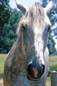
4. FLIES PLAY A ROLE IN THE TRANSMISSION OF PIGEON FEVER
C. pseudotuberculosis can survive up to two months in hay or bedding and more than eight months in soil. Exactly how the bacteria usually enter the horse’s body is still the subject of research. C. pseudotuberculosis is known to be introduced via horse-to-horse contact and by direct contact with contaminated soil. Insect bites, however, are suspected to be a primary source of disease transmission, based on Spier’s research at UC–Davis. “We went to a number of farms that were experiencing outbreaks, in several different years, and revisited those same farms in years they were not experiencing any cases. We wanted to learn more about transmission and also how these bacteria persist in the environment,” Spier says. “I worked with entomologists from UC–Davis, and we trapped flies using drift traps that capture all the insects that fly through. We also netted flies off the horses that had abscesses. When farms were having outbreaks with infected horses on the premises, we could easily find the bacteria in three different species of flies—housefly, stable fly and horn fly—and there could be some other vectors as well.”
On farms where horses had the disease, Spier’s team found that up to 20 percent of the houseflies were carrying the bacteria. “We went back to those farms on years there were no cases, and found the fly populations were negative for the bacteria,” she says. “This tells us that the reservoir is not the fly but the soil. Insect vectors can transmit the disease but the insects themselves are not the reservoir. They only assist in spreading bacteria from horse to horse.”
5. EVEN IN ENDEMIC AREAS, THE DISEASE RUNS IN CYCLES
Pigeon fever is common in many parts of California and the Southwest, but even in those regions, the number of cases increase and decrease over the years.
“Pigeon fever cases were scattered widely in our region this past year,” says Hector Gonzalez, DVM, of Bakersfield Veterinary Hospital in California. “We saw about seven horses in our practice during 2015 that had clinical presentation of pigeon fever—including one with ulcerative lymphangitis. We didn’t see as many cases in 2014. The conditions in 2015 were just right for it, with drought during the summer. At the beginning of 2016 it also seemed like there were more horses being affected, more like what we saw eight years ago.”
In Texas, the disease is even more sporadic. Cases there soared a thousandfold between 2005 and 2011 but have since tapered off. “The year before we had the big pigeon fever outbreak, we had some cases that looked like strangles, but when we cultured them they were actually C. pseudotuberculosis. We thought that was quite interesting, because we generally don’t see pigeon fever here,” says Ben Buchanan, DVM, DACVIM, DACVECC, of Brazos Valley Equine Hospital, which has multiple locations in Texas. “I haven’t seen a case here for about two years, however. We’re back to like it was before the drought. Maybe the next time we get a big drought in Texas we will see more cases again, but new infections seem to have stopped when it started raining.”
6. WARMING TRENDS MAY BE INCREASING THE RISK OF PIGEON FEVER
Climate change, which has brought hotter, more arid conditions to many regions, may be a factor in the spread of pigeon fever beyond its historic range. “Based on what we’ve seen of epidemics in Texas, Colorado and other states, it seems to increase in drought conditions,” says Spier. “The exact environmental conditions leading to epidemics are not completely understood. We know the bacteria survive very well when soils go from moist to dry conditions, which may allow the bacteria to spread more readily.
“One theory is that when weather is hot and dry horses seek shade and pass manure where they are standing in the shade,” she adds. “These organisms really thrive in soil mixed with manure. In dry conditions the soil underfoot becomes dusty and blows around. The horses are defecating, stomping their feet fighting flies, churning the soil and manure into dust, so the bacteria could potentially flourish and spread more readily in that situation.”
Dry, hot weather is likely to increase across the country as climate change takes hold. And as pigeon fever outbreaks occur, vigilance will be the key to protecting your horses. “Horse owners should closely monitor their animals,” says Gonzalez. “If a horse starts to develop abscesses in the pectoral region or inguinal0 region or ventral edema at the lower abdomen, they need to consult their veterinarian who can obtain a culture.”
This article first appeared in EQUUS issue #470, November 2016.
Save