Something is missing from the equine clinical laboratories at vet schools these days, and you’d notice it right away. What’s missing are the horses.
Between pressure to sacrifice fewer animals for educational use, new opportunities provided by computer-based education, and the development of mechanical simulation learning tools, the labs where vet students practice their manual skills and learn their anatomy are a different place.
But are the new tools as effective for learning as real animals? How are our future vets learning tactile equine-related skills and anatomy? Recent studies from the United States, Brazil, Great Britain and Austria shed light on the new wave of equine education tools that never kick or neigh or toss their heads.

Something has been happening in the veterinary world, and it’s time for news of it to leak out. Veterinary education is evolving, after remaining remarkably unchanged for its first 200 years or so.
Veterinary education was traditionally a course of study that required memorization of anatomy and the facts of medicine, surgery and pharmacology. Students learned formally by observing their professors and mentors and more informally by dissection of entire animals, organ systems or limbs. They kept journals and became proficient at illustrating anatomy and diagramming. Before widely available photography and radiography, both students and practitioners depended on their ability to record a case by notating the features of a lesion or wound, and often by drawing it.
Slaughterhouses have always been sources of study materials–and subjects–for students. Those magnificent anatomy drawings of George Stubbs were drawn from abattoir cadavers hanging from the rafters of a barn. Over 18 months of hands-on study, he peeled off layers of hide, skin, organs, and muscle to make the successive drawings until only the bones remained. Sadly, the city pound often was a source, as well.
Learning veterinary medicine was never easy, and it’s not any easier now, but what has happened is that educators know more about how people learn. They also know that the shift in gender of veterinary students from a majority of male students to a majority of female students means that there are differences in both how people learn and how they study.
The availability of computer-generated art, plastinated specimen, and preserved three-dimensional bones, limbs and organs brought the gray line drawings in textbooks alive. And more recently, innovative simulation techniques and devices have been developed to give veterinary students the opportunity to practice techniques without requiring research animals to be on hand.
But there was a political impetus for the change, as well, and an economic one.
According to the New England Anti-Vivisection Society (NEAVS), a leading humane organization opposed to experimentation with live animals, only seven medical schools and seven veterinary schools remain in the United States that practice what is known as “terminal surgeries” using animals that are euthanized for the purpose of providing educational opportunities for students.
In 2000, Tufts Cummings School of Veterinary Medicine in Massachusetts became the first veterinary college in the United States to discontinue terminal surgeries. Tufts purchases horses from a local dealer for student use in dissections, and augments its needs with horses donated by owners after treatment in the hospital; these animals now account for up to 40 percent of the horses used by students in anatomy study.
Slaughterhouses became less of an option as a source when the United States Congress technically forced horse slaughter to be done in Canada or Mexico rather than within the United States. Public health issues with handling, moving, and disposing of carcasses is also a concern. Students may have the option not to participate in dissections in some schools, if they have personal objections to the practice.
On the economic side, vet schools have traditionally been associated with large land-grant universities that also had active horse breeding programs that stood stallions and sold valuable offspring into the public each year. As these programs have evolved from large-scale breeding to more management operations, fewer excess horses are available on campus. Some vet schools also maintained a herd of horses kept strictly for research or education use, but horses, even if donated, are expensive to keep fed and healthy.
Beyond surgeries for colic, orthopedic repairs or castration, vet students still need to learn to palpate mares, inject hocks, use an endoscope and float teeth. Add in the pressure of larger classe sizes at most vet schools, and the pressure has been on to develop alternative ways for vet students to practice clinical skills without needing a horse on hand.

With the availability of 3-D technology and imaging, digital teaching resources in anatomy have increased. Investments made in the new technology seemed a safe bet but some interesting new studies question how people retain what they learn when they use different types of anatomy teaching tools.
Veterinary educators have also found differences between the ways that male and female students typically absorb and retain information.
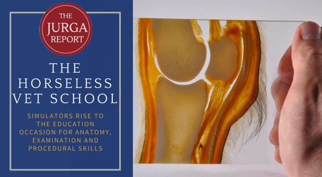
In at least one study, data showed that high tech methods are not always the most successful with students. Professor Renate Weller and a team at the Royal Veterinary College in Great Britain tested 64 vet students who volunteered to study the anatomy of the horse’s foot. The students were split into three groups, and studied either a plastic three-dimensional life-sized model of a horse’s foot, a high-tech self-guided 3-D computer presentation, or traditional textbook illustrations.
Weller reported her team’s findings in the journal Anatomical Sciences Education.
The students’ comprehension of the anatomical structures was tested by having them look at MRI images of feet. Textbook and 3-D computer program group scores were about the same, at approximately 63 percent, but when students used the plastic model that they held in their hands, the comprehension rose to 86 percent. Students also reported a more positive learning experience when using the model.

In Brazil, meanwhile, the idea was to combine 3-D thinking with real horses to teach anatomy paired with equine locomotion. Painting anatomical structures onto horses is a practice that educator Susan Harris introduced at American events long ago. British educator Gillian Higgins has used it as the basis for her popular courses, and Debranne Patillo uses it in her Equinology education system. No doubt it is used all over the world.
Equine body painting has gained sophistication and worldwide acceptance because of its success in getting people to pay attention to both the anatomy of the horse and how it moves. But how could that be proven? And is it scientifically accurate enough to be used in veterinary medicine?
In the Brazilian study, students who opted for a 90-minute painted horse session to learn anatomy over a traditional lecture hall session showed a significant improvement in their comprehension of equine anatomy and reported a positive learning experience.
What seems like a small study and finding may give legitimacy to a low-cost do-it-yourself method and encourage new expansion of the practice with some brain power applied to bigger applications.

Meanwhile, back at the Royal Veterinary College, Professor Weller put the wheels in motion for her latest anatomy education project: the definitely low-tech horse called Jarvis. Recent RVC graduate Vicky Simons was selected to paint Jarvis, using anatomy textbook references and consultations with vet school staff, and including an abdomen painted with ultrasound-guided accuracy. The result is a freestanding anatomically-accurate life-sized horse model that students will probably enjoy learning from and remember for years to come.
You can be sure that their use of it will be studied and evaluated. Such “anatomy dummies” were standard equipment in Victorian times, even to the point of have removable organs.
What about student needs when it comes to practicing their clinical procedures, such as palpating a mare or injecting a joint? The University of Vienna’s Skills Lab is a veterinary hospital whose patients are all dummies. How successful are the dummies–known as simulators in educator parlance? Two studies evaluating the simulators in Vienna have been published in the journals Theriogenology and Reproduction in Domestic Animals.
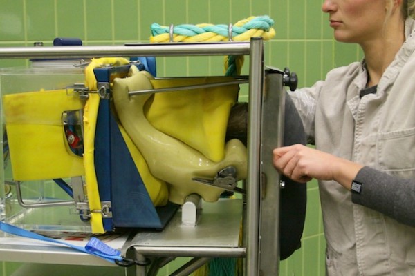
Twenty-five third-year veterinary students at Vienna participated in the study on student participation in equine reproduction examination of mares. Students were randomly allocated to three groups and taught palpation and ultrasonography of the equine genital tract in different ways. Group 1 trained four times on the simulator, consisting of a plastic box shaped like the hind end of a horse with exchangeable rubber genital organs. Group 2 students were trained four times on teaching horses whereas group 3 had only one training session on live mares.
Two weeks after the last training session, the researchers tested the learning outcomes. All students were asked to examine a mare and to make a diagnosis. Students who had trained four times on horses scored best with regard to a correct diagnosis. Students who had been trained only once on live horses had the worst results and those trained solely on the simulator scored in between the two other groups. With regard to ultrasonography of the genital tract, there were no major differences between groups.
“Simulator-based training prepares the students very efficiently for diagnostic procedures on live horses,” explains Christina Nagel, principal investigator of the study. “Simulators are, however, not only an additional teaching tool for our students but also a contribution to animal welfare. Only when students have successfully finished the simulator-based training course are they allowed to perform the same diagnostic procedures in animals.”
Students perceived that the examinations were stressful for the horses. Part of the study involved testing the students’ own stress levels by measuring their heart rates and salivary cortisol levels; students who had prepared for the live exams by practicing on the simulators had lower signs of stress.
Simulators are finding their way into many areas of the horse world. Students at the British Racing School learn to fall off horses by landing on mats after tumbling off mechanical horses. Dressage riders study animations of their tests, and eventers now benefit from course walks that are like video games. Mechanical chairs simulating the motion of a horse have been found to be helpful in physical therapy for certain handicaps.
Homemade aids teach people to braid manes and tails that aren’t attached to horses.
The possibilities for simulators in equine education are limitless. Without the need to transport horses to a workshop, costs decrease and the choice of a venue increase. Humans can be taught to evacuate horses from a fire, give inoculations, load horses on trailers and install fences by using simulators.
Most of all, a wider use of simulators can remove the barriers from people’s perceptions that learning is somehow difficult, complicated, expensive or that it requires travel or assets that may not be available. Simulators could move the horse world to a more open place where everyone is learning something, or else figuring out a creative way to teach it.
To learn more, the studies mentioned in this article can be found in the sources below:
“Let’s Get Physical”: Advantages of a physical model over 3D computer models and textbooks in learning imaging anatomy. Preece D, Williams S B, Lam R, and Weller R. (2013) Anatomical Sciences Education, 6: 216–224.
Acceptance of the bodypainting as supportive method to learn the surface locomotor apparatus anatomy of the horse. Senos R, Ribeiro MS, Martins Kde S, Pereira LV, Mattos MF, Kfoury Júnior JR, Rodrigues MR. (2015) Folia Morphologica 2015;74(4):503-7.
Three-dimensional anatomy. (news article) Simons, V Veterinary Record 2015 177: i-ii
Teaching of diagnostic skills in equine gynaecology: Simulator-based training versus schooling on live horses. Nagel, C; Ille, N; Aurich, C; Aurich, J Theriogenology, Volume 84, Issue 7, 15 October 2015, Pages 1088-1095
Stress Response of Veterinary Students to Gynecological Examination of Horse Mares – Effects of Simulator‐Based and Animal‐Based Training Nagel, C; Ille, N; Erber, R; Aurich, C; Aurich, J Reproduction in Domestic Animals, Volume 50, Number 5 pp. 866-871(6)
Also:
Elegant and Exact: George Stubbs’s The Anatomy of the Horse (Metropolitan Museum of Fine Art blog)