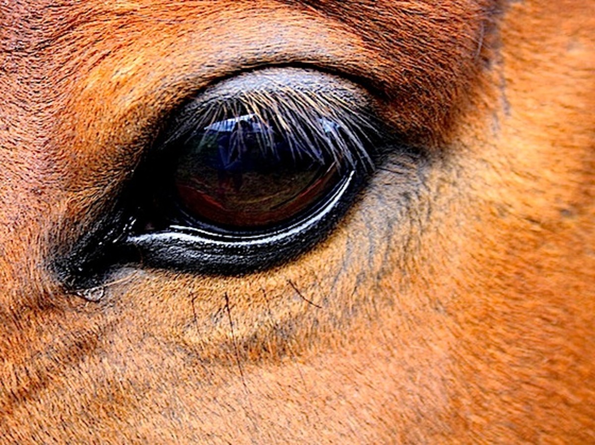
Horses face high risks of developing eye problems, and Cornell clinicians have developed a new way to detect and diagnose them more safely and quickly than before. Published online in January 2014 in the journal Veterinary Ophthalmology, their findings are the first to show how horses with microscopic foreign objects in their eyes can benefit from in vivo corneal confocal microscopy, a human medicine technique that let doctors take pictures of living eyes in microscopic detail without a scratch.
After veterinary ophthalmologist Eric Ledbetter, DVM, DACVO began adapting the technique in feline and canine patients at Cornell University Hospital for Animals, he discovered two new infectious diseases of the eye that had never been described before.
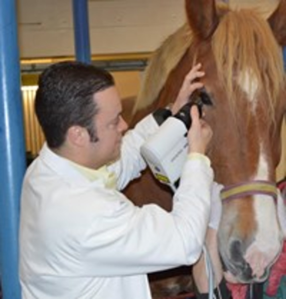
Using an in vivo corneal confocal microscope with a focal depth of 1.5mm he adapted for use on horses, Ledbetter demonstrated its ability to repeatedly examine and take images all the way through a horse’s 1 mm-thick cornea— the eye’s first line of defense and a frequent site of injury and infection.
Unlike traditional methods of eye imaging, confocal microscopy gets immediate results without needing a biopsy or any other kind of surgery.
Ledbetter has used it in clinics to help find and characterize tumors, scratches, foreign bodies, infections, immune-mediated ocular diseases, and other eye problems. He has published past articles describing the normal features of the equine cornea as viewed using in vivo confocal microscopy and the use of the technique to diagnose horses with fungal keratitis (fungal infections of the cornea).
Ledbetter then expanded to other species. He became the first to use the technique to examine horses, pioneering a clinical research program to develop and validate non-invasive eye imaging in a species particularly poised to benefit from it.
“Horses have very prominent eyes and live in environments that put their eyes at risk of trauma,” said Ledbetter. “They frequently have diseases of the ocular surface and other eye problems for which corneal confocal microscopy will be particularly useful. For example, horses frequently get fungal infections of the cornea. This has traditionally been a hard problem to diagnose — regular culturing methods of diagnosing fungal infections can take 10 to 14 days for results to come back, creating long treatment delays.”
Using an in vivo corneal confocal microscope with a focal depth of 1.5mm he adapted for use on horses, Ledbetter demonstrated its ability to repeatedly examine and take images all the way through a horse’s 1 mm-thick cornea— the eye’s first line of defense and a frequent site of injury and infection.
Unlike traditional methods of eye imaging, confocal microscopy gets immediate results without needing a biopsy or any other kind of surgery.
Ledbetter has used it in clinics to help find and characterize tumors, scratches, foreign bodies, infections, immune-mediated ocular diseases, and other eye problems. He has published past articles describing the normal features of the equine cornea as viewed using in vivo confocal microscopy and the use of the technique to diagnose horses with fungal keratitis (fungal infections of the cornea).
But when it came to identifying microscopic corneal foreign bodies using the new technique, he first had to validate its accuracy. By collecting images of horses’ eyes with foreign bodies and comparing them to results from traditional biopsy-based diagnostic methods like cytology and histopathology, he validated confocal microscopy as a quicker non-invasive technique to image and diagnose this eye problem accurately.
“With any new technology like this, you can take a lot of images but no one knows for sure what the images represent,” said Dr. Ledbetter. “You might have an idea, but in the research stage you have to validate every hunch. By concurrently using both new and preexisting techniques, we compiled and published evidence that findings match. This paves the way for veterinarians to definitively diagnose eye diseases in horses with only this new technology, minimizing impact on the eye and saving time to get patients treatment faster.”
To learn more:
Ledbetter, E. C., Irby, N. L. and Schaefer, D. M. W. (2014), In vivo confocal microscopy of corneal microscopic foreign bodies in horses, published in Veterinary Ophthalmology.