
If injuries in racehorses can be predicted, is there a greater chance they can be prevented?
Yes, reveals the latest free podcast in the Equine Veterinary Journal (EVJ) audio collection.
In a 45-minute orthopedic-themed broadcast, Tim Parkin, BSc,BVSc, PhD, DipECVPH, FHEA, MRCVS discusses the advantages of early MRI detection of bone changes known to precede catastrophic limb fracture. He is joined by Florida equine practitioner Sarah Plevin, BVMS, MRCVS, CVA, DABVP, DACVSMR, who talks about the possible relationship between sesamoiditis, subclinical suspensory ligament branch change and suspensory ligament branch injury in yearling Thoroughbreds.

Fracture of the lateral condyle of the third metacarpus (MC3, or “cannon bone”) is the most common reason for euthanasia on the racecourse, while suspensory branch ligament injury is a frequent stumbling block for yearling Thoroughbreds in the initial months of training.
In the podcast, Parkin and Plevin both support and explain the potential advantages of early intervention to prevent injury and maintain racing performance.
Parkin, from the Glasgow University Veterinary School in Scotland, explores the question, “Can we use subchondral bone thickness on high-field magnetic resonance images (MRI) to identify Thoroughbred racehorses at risk of catastrophic lateral condylar fracture?” (1) His study, which assessed the bone-level risk factors for fracture in racehorses, is part of a long-standing program of research to prevent racehorse injury, funded by the Horserace Betting Levy Board (HBLB) in Great Britain.
In the study, 191 cannon bones from 96 horses with (47) and without (49) lateral condylar fracture were subjected to magnetic resonance (MR) imaging. Greater depth of dense subchondral/trabecular bone in the palmar half of the lateral parasagittal groove of distal MC3 (end of cannon bone at the fetlock joint) was associated with an increased likelihood of being from a horse that had sustained a fracture.
The identification of such markers for pre-fracture change that can be detected using MRI suggests that reliable screening methods for fracture risk may be developed. The routine use of such screening programs would enable interventions, such as alterations to training regimens based on known risk factors for lateral condylar fracture that may reduce the likelihood of fracture in susceptible horses.
Plevin, of Florida Equine Veterinary Associates in Ocala, Florida, continues the podcast with findings on associations between sesamoiditis, subclinical ultrasonographic suspensory ligament branch change and subsequent clinical injury in yearling Thoroughbreds. (2)
Sesamoiditis is a common radiological finding in yearling Thoroughbreds. The condition is believed to be associated with suspensory ligament branch injury (SLBI), which is known to affect racing performance.
Plevin’s observational study evaluated 50 untrained yearling Thoroughbreds at a training center. The results identified a significant relationship between grades of sesamoiditis, subclinical suspensory ligament branch change (SSLBC) and subsequent suspensory ligament branch injury (SLBI). The associations highlight the importance of ultrasonographic examination of suspensory ligament branches in horses with significant grades of sesamoiditis.
Ultimately, these findings could allow more accurate prognoses to be made regarding the development of SLBI and provide opportunity for intervention and prevention of such injury.
Professor Celia Marr, Editor of the Equine Veterinary Journal said: “Major advances in diagnostic imaging mean that we are increasingly able to deploy predictive science in order to prevent serious injury. Important studies such as these and others supported by the HBLB are giving us real potential to rule out horses at risk and thus reduce significantly some of the welfare concerns in the horseracing world.”
The free EVJ podcast archive is available at: http://apple.co/1KnWqX7 .
References in the Equine Veterinary Journal to support the podcast; the first paper is open for all to view, while the second requires a subscription, web access or purchase to view:
(1) Can we use subchondral bone thickness on high-field magnetic resonance images to identify Thoroughbred racehorses at risk of catastrophic lateral condylar fracture? C. A. Tranquille, R. C. Murray, T. D. H. Parkin DOI: 10.1111/evj.12574
(2) Association between sesamoiditis, subclinical ultrasonographic suspensory ligament branch change and subsequent clinical injury in yearling Thoroughbreds. S. Plevin, J. McLellan, T. O’Keeffe DOI: 10.1111/evj.12497
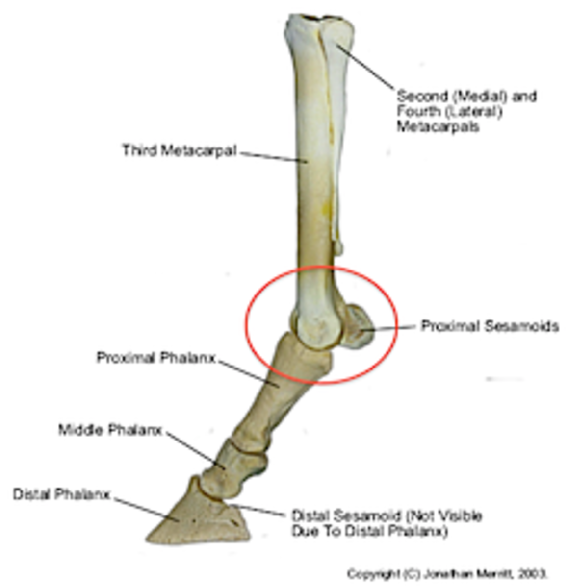
Skeletal anatomy illustration courtesy of Jonathan Merritt, PhD, University of Melbourne. Dr Merritt kindly published the illustration under a Creative Commons,Attribution-Share Alike 3.0 Unportedlicense.

Muscular anatomy image at top courtesy of Wellcome Images library, Creative Commons Attribution International 4.0 license.


