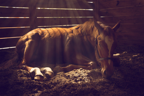
Nantucket had been with his new owners for only one month when the colic struck. The 5-year-old Thoroughbred-cross gelding was purchased as a potential show hunter, but he hadn’t yet made it to his first competition when his new family found him one morning pawing, restless and uninterested in his feed.
The local veterinarian came out right away, and he agreed with the family’s assessment that the colic was still mild: The gelding was far from the frantic, sweaty state that means acute gut pain. Nevertheless, the veterinarian began a full diagnostic workup.
A check of vital signs revealed only a slightly elevated heart rate. Next, the veterinarian passed a nasogastric tube to check for a buildup of fluid in the gelding’s stomach, but only a small amount flowed from the tube into the waiting bucket, which was an encouraging sign. The rectal palpation, however, yielded some troublesome findings. Nantucket’s large colon felt like an inflated beach ball—a sign of gas buildup.
The veterinarian gave the gelding a dose of Banamine. Often, mild colics will subside after the painkiller is administered because the horse is able to relax, which allows gas to move and normal gut activity to resume. Hoping for an easy resolution, the veterinarian told Nantucket’s owners to keep a close eye on the situation and to call immediately if the gelding took a turn for the worse.
Nantucket didn’t get any worse over the course of the day, but he didn’t get any better, either. That evening the veterinarian returned and repeated the rectal palpation. The distention of the colon was growing larger, and this time, he found that the horse’s spleen had shifted—it was further forward toward the midline of the body than normal, and the colon was now traveling between the spleen and body wall up toward the nephrosplenic space. This last clue pointed toward a specific cause of Nantucket’s gut pain: nephrosplenic entrapment of the large colon.
Caught in a trap
Nephrosplenic entrapment occurs when the large colon slips upward over the spleen and gets caught on the nephrosplenic ligament, which connects the spleen to the left kidney. The ligament is located near the abdominal wall of the left flank, toward the top of the body. When the colon gets trapped against this ligament, gas and feed cannot move easily through it, causing a backup that leads to colic pain.
Why this condition occurs is not known. “There are a few theories,” says Clarisa Krueger, DVM, of Elgin Veterinary Hospital in Elgin, Texas. “There are nerve plexus in the colon, responsible for normal motility, and if those plexus fire wrong you can get some abnormal motility of the large colon, which allows more gas to build up and the colon to shift in position. There’s also a notion out there among horsepeople that it’s caused by rolling, which cannot be ruled out as having a role in colic, but I’m pretty sure that’s not true. We’d have horses trapping their colons on a daily basis if rolling caused it.”
Nevertheless, Nantucket showed the two classic signs that point to this diagnosis: an “inflated” colon caused by the backed up gases and food matter, combined with the shift in the position of the large colon and spleen.
The local veterinarian went over treatment options with the gelding’s owners. If Nantucket had been showing signs of severe pain—such as thrashing, sweating or a highly elevated heart rate—the first recommendation would have been immediate transport to a clinic for emergency surgery.
However, because Nantucket’s pain was still fairly mild, the veterinarian suggested trying a more conservative treatment first: The gelding would be injected with the drug phenylephrine, which reduces the size of the spleen by triggering it to release all of its stored red blood cells. Then Nantucket would be jogged on a longe line for about 30 minutes. The goal of the gentle exercise is to “jostle” the colon off the ligament and back into its proper position.
The “medication-and-jog” approach is successful in a majority of cases—63.2 percent, according to one 2013 study—yet it does carry some risks. If the colon is very heavy with backed-up feed, the jarring could damage the intestinal wall. But Nantucket’s case didn’t seem to be that precarious.
With the owners’ consent, the veterinarian proceeded with the conservative treatment. Normally, phenylephrine and exercise will relieve an entrapment quickly, and the horse will appear more comfortable instantly. Nantucket, however, still appeared colicky an hour later.
The next step, the owners agreed, was to take Nantucket to Elgin Veterinary Hospital for more intensive treatment and possibly surgery.
Choices to make
“I remember he looked agitated but not awful,” says Krueger, when she met Nantucket at the trailer in Elgin. “A little sweaty and bloated and like he’d not had any rest, but with steady vital signs, and still holding it together mentally.”
A full workup, including an ultrasound examination, confirmed the local veterinarian’s diagnosis: A portion of Nantucket’s colon was trapped between his kidney and spleen. Krueger discussed the next steps with Nantucket’s owners. “At this point there are a few options,” she says. “We could continue with medical therapy using more painkillers, fluids and perhaps trying phenylephrine again.”
Another approach is to place the horse under general anesthesia in a padded room and then roll and jostle his body manually in an attempt to dislodge the colon. This procedure may resolve the entrapment, but placing a horse under general anesthesia carries other risks, including injury as a horse scrambles back to his feet upon waking, and physically rolling a large, unconscious horse can be difficult.
A third option is a standing procedure. The correction for a nephrosplenic entrapment is the only colic surgery that can be performed with the horse still conscious and under heavy sedation. “Typically, in colic surgery, you need the horse to be under general anesthesia and positioned on his back so you can make an incision along the midline to access the source of the problem,” says Krueger. “But in these entrapments the trouble spot is near the abdominal wall, high up on the left flank.”
Another advantage of this type of surgery is that it requires only a small incision that is under much less tensile stress than one in the midline of the belly. This factor dramatically shortens recovery time and reduces the risks of incision-related complications.
After discussing their options with Krueger, Nantucket’s owners agreed to the standing surgical approach.
Operating by feel
Nantucket was sedated and placed in stocks that would help to stabilize him if he became unsteady on his feet. His left flank was then clipped and scrubbed with Betadine and his body covered in sterile drapes.
“You start this procedure by making a 15- to 20-centimeter [six- to eight-inch] incision on the left flank, between the last rib and hip,” says Krueger. “It’s not very large, just long enough for a surgeon’s arm to fit in.”
In a normal horse, the spleen is immediately accessible through this incision, but in cases of nephrosplenic entrapment, the spleen is pushed out of position and a surgeon will first encounter the displaced large colon.
Krueger slowly pushed her arm into the incision, carefully feeling her way around to orient herself to the placement of organs. “If you feel multiple issues or even something you can’t quite identify, you don’t want to continue with the horse standing,” she says. “At that point, you backtrack to go to general anesthesia.”
Feeling confident that the entrapment was Nantucket’s only issue, Krueger cupped her hand underneath the colon and gently pressed the spleen toward the floor of the abdomen. “This allowed me to lift the colon up over the spleen, toward the outside of the horse and then let it drop,” she says. “The nephrosplenic ligament acts like a coat hook, and you have to lift the colon up and off the hook.”
Freeing the colon was only the first step. Still working entirely by feel, Krueger palpated the intestine to estimate the texture of the contents. “Sometimes if the horse has been backed up for a while, the gut will be full of really firm or dry ingesta that won’t move easily on its own,” she says. “In those cases, we will do a standing enterotomy, where you flush the area with fluids.” Nantucket’s colon, however, was filled with soft, doughy material that was likely to move on its own.
Continuing her check, Krueger reached down toward the floor of the abdomen and felt Nantucket’s pelvic flexure, the portion of the large colon that folds back on itself in a hairpin turn. “This turn is supposed to be positioned pointing toward the back of the horse,” she says. “In this guy it was out of place, pointing toward the front. It’s technically called ‘pelvic flexure retroflexion.’”
Whether this retroflexion was the cause or a result of the entrapment was unclear, but at this point, it didn’t matter. Krueger began the careful and difficult work of repositioning the colon. “You are working with only one arm, deep within the horse, and you can’t see anything,” she says. “You have to move the colon very carefully, inch by inch. It’s physically very demanding, and we generally take turns working because it’s very exhausting.” The entire surgery took about an hour, and Nantucket stood quietly throughout.
Once all the organs were back in their proper places, Krueger closed the incision on Nantucket’s flank, and as the sedation wore off, the gelding gradually became more alert. It was clear he was no longer in pain. “You can tell almost immediately when this procedure works,” she says. “His heart rate was normal, he wasn’t sweaty or anxious, he had good gut sounds and he was looking for something to eat.” Unfortunately for Nantucket, it would be another 24 hours before he would be allowed to have hay.
“Recovery is much easier from this surgery than a typical colic surgery,” says Krueger. “He just needed two weeks of stall rest and walking, followed by a week of light turnout before he could be ridden again. If he had had an incision on his midline, he would have been out of work for months to minimize pressure on it and allow it to heal.”
Twenty-four hours after surgery, Nantucket developed a slight fever, which isn’t unusual. “He was on anti-biotics at the time, and it was short lived,” Krueger says. “He went home two days after surgery.” The fever returned a week later, which prompted some concern, a round of bloodwork and more antibiotics, but that one also resolved quickly. “We never found out what caused that second fever, but ultimately it wasn’t a big problem,” Krueger adds. Ten days after surgery, a member of the veterinary team made a final visit to Nantucket’s farm to remove the incision’s staples.
Recently, Krueger wrote a paper for a veterinary journal on standing surgical correction of nephrosplenic entrapments. As part of her research, she contacted Nantucket’s owners to find out how the gelding was doing two years after his ordeal: “He’s doing fantastic,” she says. “He’s a hunter/jumper and doing everything the family had intended. He’s had no repeat colic episodes and seems to be an all-around great guy.”
This article first appeared in EQUUS issue #449, February 2015.


