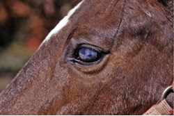
Spark’s right eye was not a pretty sight. The 7-year-old Quarter Horse stallion had tangled with something in his paddock —no one was certain what—and he arrived at the University of Saskatchewan’s veterinary clinic late Saturday evening with a large laceration on his right foreleg and a gruesome-looking eye: The entire globe was cloudy, and a small piece of brown tissue from the interior was projecting out through the surface.
However, veterinary ophthalmologist Lynne Sandmeyer, DVM, was guardedly optimistic when she first met and examined Spark. “His eye did look alarming,” she says, “and his owners were understandably very worried. But they had brought him in immediately, which increased his chances of a good outcome.”
After a general physical exam, which showed Spark to be healthy apart from his injuries, the veterinary team tended to the laceration on the stallion’s foreleg first. Although the wound was large, it hadn’t affected any tendons or joints, and the horse wasn’t significantly lame.
“He didn’t need any sutures, but we did clean and debride the wound thoroughly,” says Sandmeyer. “We also applied a bandage that he’d need for the next few days.”
Then the team turned their attention to Spark’s eye. “Looking just with a penlight, we could see there was a clean, full-thickness laceration on his cornea, the outer surface of the eyeball,” says Sandmeyer. “It was the type of cut that’s made from something very sharp, like a wire.” She also noted that the brown tissue projecting from the laceration was a piece of the horse’s iris—the pigmented structure within the eye that controls the size of the pupil by constricting and expanding.
But as shocking as it looked, the fact that Spark’s iris was projecting out through the front of his eye wasn’t unusual—and it could even be considered a good thing.
“As soon as a horse sustains a full-thickness cut through the cornea, aqueous fluid within the eye begins rushing out, causing an immediate drop in pressure,” says Sandmeyer. “As the pressure drops, things are sucked out through the wound. The iris, which normally sits in front of the lens, gets pulled forward into the laceration. This ends up ‘plugging’ the leak, as it were, and actually works to stop the fluid loss.”
Trauma to the eye also triggers an influx of inflammatory-related substances, one of which is the sticky protein called fibrin. “Fibrin collects around the laceration and the displaced iris to form a tight seal around the wound,” says Sandmeyer. “This all happens in a few minutes after such an injury and is a pretty effective way of dealing with the immediate problem of aqueous fluid loss.”
Sandmeyer continued the ocular exam. She saw no response to the “menace” test, meaning Sparks didn’t reflexively blink or attempt to avoid a hand coming toward his eye. This suggested that he wasn’t seeing clearly, if at all, out of his right eye.
“His lack of vision could have been due to a few things,” says Sandmeyer. “For starters, his eye was cloudy. Normally, the fluid in the eye is crystal clear, but when proteins accumulate with inflammation, that fluid starts to become cloudy, and the horse simply can’t see through it.”
Other possibilities included a damaged lens or retina. In a healthy eye, these internal structures can be seen with a tool called a slit lamp, essentially a hand-held microscope with a light source. But the cloudiness in Spark’s eye made it impossible for Sandmeyer to see these structures: “We’d decided to get the inflammation under control and reassess him Monday morning.”
A second look
In addition to antibiotic eye drops, systemic antibiotics and a tetanus booster, Spark was started on intravenous Banamine along with a topical nonsteroidal medication and atropine applied directly to his injured eye every six hours. “Atropine helps reduce pain by preventing muscle spasm in the eye. It also dilates the pupil to prevent adhesions of the iris to the lens,” says Sandmeyer. “When an eye is full of inflammatory proteins and the iris is damaged, it can start to stick to the lens, which sits behind it. If you can keep the pupil from constricting you can prevent that. That’s one of the reasons it’s so important to get treatment right away for eye injuries: The longer inflammation goes unchecked, the more potentially permanent damage it will cause.”
After two days, the medications had resolved some of the inflammation in Spark’s eye and Sandmeyer could re-evaluate the ocular exam. The eye was still too cloudy to see the internal structures adequately, however. Shining a light into the right eye caused both of Spark’s pupils to constrict—an indication that the retina was likely functioning. Next, Sandmeyer placed an ultrasound probe directly onto the eye to look for any abnormalities, including foreign objects that might have been embedded in it. Everything looked normal.
“We also performed electroretinography, a test that measures the electrical response of cells in the retina,” says Sandmeyer. “Those results were normal as well.” All the structures that support vision appeared to be intact and functioning, and indeed, Spark’s prognosis for regaining vision was good.
Sandmeyer presented Spark’s owners with three treatment options. “We could have removed the eye entirely,” says Sandmeyer. “But that seemed a little extreme given that the potential for return of vision was possible.” Another option was to simply continue medicating the eye with topical medications hoping that the laceration would heal on its own—but the effect that the adhesion of the iris within the wound might have on the ability of it to heal and the future of Spark’s vision because of this were impossible to predict. The third option was surgery to remove the protruding iris and repair the laceration. This would most likely minimize scarring and produce the best outcome for Spark’s vision. The owners opted for surgery.
A finely focused operation
Spark was placed under general anesthesia for the procedure. “Ocular surgery is delicate work in a very small area,” says Sandmeyer, “so we use a microscope focused directly on the eye to see what we are doing.”
Sandmeyer opened the laceration with a scalpel and pared off the ragged wound edges. She also removed the portion of the iris that had been projecting out of the globe. “The rest of the iris was still attached and healthy, so we just let that float back into the eye,” she says. “He’ll always be missing a tiny piece of it, but that won’t affect how the rest of it functions; his pupil will still dilate and constrict.”
Sandmeyer did a second vision check of the lens to ensure it wasn’t ruptured or otherwise damaged. “If it had been, we could have removed it, leaving him really farsighted, or put in a prosthetic lens.”
Next, she flushed out the cloudy aqueous fluid and replaced it with a balanced salt solution. “The body generates eye fluid rapidly, and so within a few hours of surgery, the salt solution is replaced with healthy aqueous fluid,” she says.
Sandmeyer stitched the laceration closed with tiny sutures and created a conjunctival graft to protect the area. “We create a small stalk of tissue, from the conjunctiva around the eye,” she explains. “One end is still attached to the blood supply, but the other is sewn over the wound. This flap protects the area.”
Spark recovered uneventfully from the anesthesia and seemed to be able to see clearly right away. The next day the stallion was sent home with detailed instructions for three weeks of medication. “Medicating eye injuries can be very time-consuming and difficult,” says Sandmeyer. “He was getting multiple medications in his eye four times a day in the beginning. Eventually, horses start to object, and it can become quite the battle.”
To make the process easier, Spark was outfitted with an ocular lavage system, a narrow tubing placed under the eyelid and secured to his mane. Then, his medications could be injected into the end of the tube to be delivered directly into his eye.
After three weeks, Spark was returned to the clinic for a follow-up exam. “He healed really well,” says Sandmeyer. “The eye was clear and all our tests indicated his vision was functionally normal. There’s a lot that could have gone wrong in those three weeks; infection could have developed, and some horses end up damaging the eye again if they rub it on something. But his eye looked great.”
Conjunctival grafts, like Spark’s, are left in place indefinitely but do not interfere significantly with vision. “He has to see around it, but it’s very tiny,” says Sandmeyer. “And even if the graft wasn’t there, he’d still have the scar on that area blocking his vision.”
Despite how serious the injury had appeared at first, Spark’s positive outcome isn’t unusual, says Sandmeyer: “I know eye injuries can look really bad, but if you get help right away and are able to control the inflammation you’d be surprised just how well they can do.”
This article first appeared in EQUUS issue #446, November 2014.


